 by
by 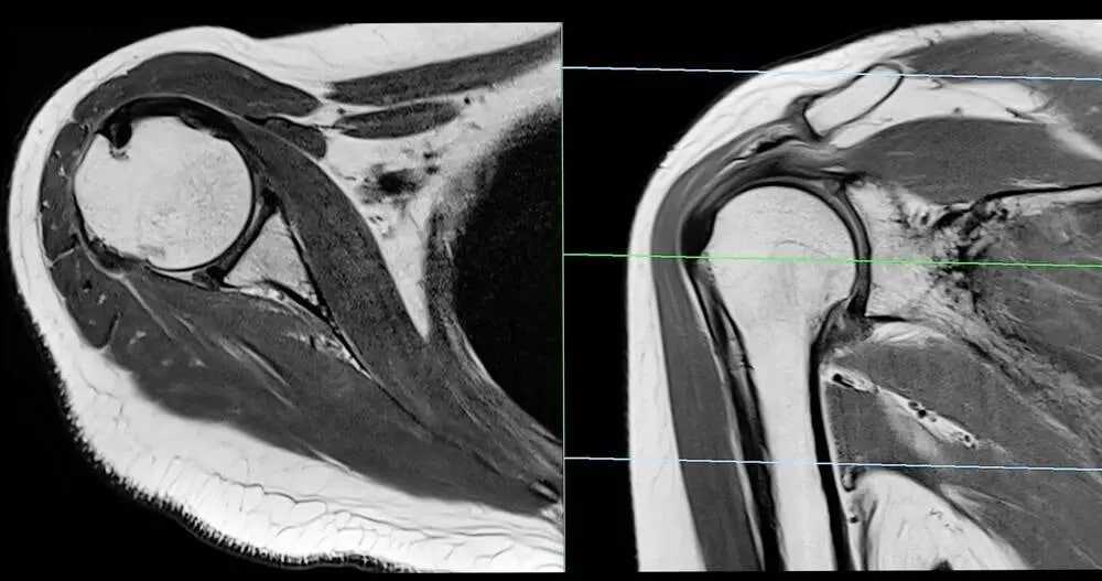
Magnetic Resonance Imaging, widely known as MRI, has become a tool in modern diagnostic medicine. Particularly this is used for identifying soft tissue injuries. Unlike X-rays, MRI provides layers of detail about muscles, tendons, and ligaments. This aids in the diagnosis of conditions that might otherwise remain hidden. Its non-invasive nature and ability to deliver highly detailed images have made it a choice for medical professionals in managing soft tissue conditions.
How Does an MRI Work?
MRI relies on powerful magnetic fields and radio waves to generate images of the body’s interior. This approach makes it particularly effective for soft tissue imaging since it captures the contrast between different types of tissues. With the ability to highlight fine details, MRI scans can detect tears, swelling, or abnormalities within soft tissues.
The process involves placing the patient within a cylinder-shaped scanner that takes cross-sectional images of the area under observation. These images allow physicians to pinpoint the nature and location of an injury more accurately than other imaging methods. This accuracy is especially relevant in sports-related injuries.
What are Common Soft Tissue Conditions Evaluated?
Soft tissue injuries span a range of conditions, many of which benefit from the clarity provided by MRI imaging. Ligament injuries, such as ACL or MCL tears in the knee, are frequently assessed using this technology. Tendon conditions are often diagnosed through MRI scans. MRI is also used for more complex conditions affecting soft tissues, including tumors or cysts. By offering clear images of soft tissue masses, this helps in detection and evaluation, guiding physicians in determining whether further treatment is necessary. Detailed imaging is valuable not only for identifying initial issues but also for monitoring recovery, as it allows tracking of tissue repair over time.
What are Advantages of Using MRI?
One of the key advantages of MRI is its ability to provide high-resolution images without relying on radiation. This makes it a safer option for patients requiring multiple scans over time. MRI is particularly well-suited for obtaining images of soft tissues that cannot be viewed effectively with X-rays or ultrasounds.
MRI also supports detailed, multi-plane views of the affected area. These comprehensive perspectives give medical professionals a complete understanding of the injury. This level of detail reduces the margin of error in diagnosing soft tissue injuries, ultimately aiding in the selection of the most appropriate therapy or intervention.
When is an MRI Recommended?
Healthcare providers may recommend an MRI when other imaging techniques fail to provide sufficient information about a soft tissue injury. Patients experiencing persistent pain, swelling, or limited mobility in an affected area often undergo this type of imaging. The versatility is particularly evident in its application across a variety of conditions. Patients considering an MRI should consult with their medical professional to help make sure that the scan aligns with their specific condition and symptoms. While MRI is a valuable diagnostic tool, it is most effective as part of a comprehensive medical assessment.
Take the Next Step Toward More Precise Diagnosis
For individuals who suspect they might have a soft tissue injury, discussing imaging options with a healthcare provider is a practical first step in addressing the issue. Whether you are managing a sports injury or coping with another condition affecting muscles, tendons, or ligaments, early and accurate diagnosis can support better treatment outcomes. MRI offers a reliable option to investigate and monitor these injuries, providing vital clarity to patients and medical professionals alike.